نسبة نجاح عملية المياه البيضاء

Success Rate of Cataract Surgery

تشير المياه البيضاء إلى حالة تصيب عدسة العين وتؤدي إلى تغيرات في شفافيتها. تظهر العدسة غائمة بشكل تدريجي، مما يشبه النظر إلى سطح زجاجي مشوش أو مغطى بالثلج. يؤدي تكون المياه البيضاء إلى منع مرور الضوء من خلال العدسة وتشوش الرؤية، مما يتسبب في صعوبة قراءة النصوص وغيرها من المشاكل البصرية. يمكن أن تصيب المياه البيضاء الأشخاص في أي عمر وتؤثر على إحدى العينين أو كلتيهما.


نسبة نجاح عملية إزالة المياه البيضاء:

تبلغ نسبة نجاح عملية إزالة المياه البيضاء (Cataract surgery) على العين نسبة تتراوح عادة بين 95% إلى 99%. يمكن أن تتفاوت هذه النسبة بناءً على العوامل المختلفة المتداخلة في العملية.

معلومات تهمك تكلفة عملية المياه البيضاء بالليزر لعام 2023

العوامل المساهمة في زيادة نسبة نجاح عملية إزالة المياه البيضاء:

تعد عدة عوامل مهمة لزيادة نسبة نجاح عملية إزالة المياه البيضاء. من هذه العوامل:

  1. خبرة الجراح: تلعب خبرة الجراح دورًا مهمًا في زيادة نسبة نجاح العملية. كلما كان للجراح خبرة ومهارة في إجراء هذه العملية، زادت فرص النجاح.
  2. سمعة الجراح: يعتبر سمعة الجراح وسجله الناجح من العمليات السابقة أحد العوامل المؤثرة في نسبة نجاح العملية.
  3. التقنيات والتجهيزات: استخدام التقنيات والأدوات المتطورة في عملية إزالة المياه البيضاء يمكن أن يحسن من نسبة النجاح.
  4. الحالة الصحية للمريض: تلعب حالة صحة المريض دورًا في النجاح، حيث أن الحالات الصحية المعقدة قد تزيد من مخاطر العملية.
  5. المرافق المستخدمة: اختيار مستشفى أو مركز يكون مجهزًا ومؤهلاً لإجراء عملية إزالة المياه البيضاء يعزز النجاح.
  6. الامتثال للتعليمات بعد العملية: يلعب اتباع المريض لتعليمات ما بعد العملية دورًا هامًا في الحصول على نتائج ناجحة وتعافٍ جيد.

مع ذلك، يجب أن يتم تقييم كل حالة بشكل فردي، ويجب استشارة الجراح المختص لتحديد النسبة المتوقعة لنجاح لعملية بناءً على العوامل الفردية والظروف المحيطة.

إحجز موعدك الأن

يجب استشارة الطبيب في الحالات التالية:

  • إذا لاحظت أي تغيرات في الرؤية مثل ظهور هالات حول الأضواء أو صعوبة في القيادة ليلاً بسبب وهج المصابيح الأمامية.
  • إذا شعرت بألم في العين أو تعاني من صداع مفاجئ.


هل المياه البيضاء تسبب العمى؟

في حالة عدم معالجة حالة الماء الأبيض وعدم استبدال العدسة المتضررة، فإن الحالة قد تتطور وتؤدي إلى فقدان البصر. ومع ذلك، بعد إجراء الجراحة واستبدال العدسة المتضررة، يعود الرؤية إلى حالتها الطبيعية.

هل يجب أن يكون المريض في مرحلة متقدمة لإجراء عملية العلاج الجراحي؟

بفضل تطور جراحة العيون المجهرية واستخدام تقنيات تفتيت العدسة (الفاكو)، لم يعد من الضروري الانتظار حتى تصل حالة الماء الأبيض إلى مرحلة متقدمة. يمكن إجراء العملية في أي مرحلة من تطور الماء الأبيض.

من المهم مراجعة طبيب العيون لتقييم الحالة الفردية واستشارته حول الوقت المناسب لإجراء الجراحة والخيارات المناسبة للعلاج.

إحجز موعدك الأن


طرق علاج المياه البيضاء على العين

عملية المياه البيضاء بالغرز الجراحية

في هذه العملية يتم الاعتماد على كفاءة و مهارة الطبيب الجراح كلياً. حيث يقوم بعمل شق جراحي طولاني بجوار قرنية العين مباشرة للوصول إلى العدسة العاتمة و إزالتها كما هي. ثم يتم وضع عدسة جديدة وإغلاق الجرح بواسطة الخيوط الجراحية أو الغرسات. وعلى الرغم من أن هذه العملية من أقدم الإجراءات لعلاج المياه البيضاء. لكن قد يتم اللجوء إليها حتى الآن في حال كانت المياه البيضاء قديمة جداً أو متحجرة. وبالتالي يصعب تفتيتها بأشعة الفيمتو ليزر أو الموجات فوق الصوتية.

إجراء عملية المياه البيضاء بالفيمتو ليزر

تستغرق هذه العملية كاملة 5 دقائق فقط و يتم فيها تخدير العين موضعياً. و من ثم يتم توجيه ليزر الفيمتو إلى داخل العين لتفتيت المياه البيضاء المتجمعة فيها وعمل فتحات دقيقة للغاية داخل القرنية. ليتم من خلالها سحب المياه البيضاء بشكل كامل. ومن ثم يتم استخدام الفيمتو ليزر مرة ثانية لتصحيح مشاكل النظر مرة أخرى.

عملية المياه البيضاء بالفاكو

في هذه الطريقة يتم القيام بفتحات جراحية دقيقة جداً داخل العين ليتم من خلالها شفط المياه البيضاء المتراكمة بتقنية الموجات فوق الصوتية التي تسمى (الفاكو). و تتميز هذه الموجات بأن لها حركة ترددية أو اهتزازية مناسبة للعين ثم يتم جلب عدسات عينية مطوية ذات جودة عالية، و تزرع من خلال الفتحات الجراحية الدقيقة في العين و التي لا تتطلب أي خيوط جراحية أو غرزات إنما تلتئم بشكل تلقائي خلال فترة قصيرة لتنتهي بذلك العملية ويستعيد المريض الرؤية بوضوح مجدداً.

إحجز موعدك الأن


هل يمكن علاج حالة المياه البيضاء دوائياً؟


“تزعم العديد من المتاجر الإلكترونية و محلات العطارة بوجود العلاج النهائي للمياه البيضاء و هو على شكل قطرات عينية”
أولاً لابد من الإشارة إلى أن العين هي أكثر عضو حساس في جسم الإنسان و بالنظر إلى تشريحها الوظيفي. فهي أكثر منطقة غنية بالتروية الدموية. ولذلك يتم الامتصاص فيها سريعاً جداً. أي أن المستحضرات التي تطبق بشكل عيني يجب أن تكون عقيمة للغاية ومصرح بها من قبل وزارة الصحة و مخصصة للعين.

  لكن المنتجات التي تباع بشكل عشوائي لا تملك أي من هذه الصفات الثلاثة و لا ننصح بتجربتها أبداً. لأنه في الواقع لا يوجد أي علاج دوائي لحالة المياه البيضاء ولا يتم شفاؤها إلا عن طريق شفط المياه البيضاء بإحدى الإجراءات و استبدال العدسة بعدسة جديدة، و هذا لا ينفي إمكانية استحداث قطرات عينية لعلاج حالة المياه البيضاء مستقبلاً، لكن في وقتنا الحالي لا يوجد أي منتج كهذا.

يعد البروفيسور أحمد المعتصم خبير في عملية المياه البيضاء

لدى الدكتور أحمد المعتصم افضل دكتور مياه بيضاء خبرة هائلة ومهارة مثبتة في مجال علاج مياه البيضاء. يتمتع بخبرة واسعة في تشخيص ومعالجة حالات مياه البيضاء باستخدام أحدث التقنيات والفحوصات الدقيقة المعتمدة عالميًا. يتفهم تمامًا التحديات والمتطلبات الفردية لكل مريض ويعمل بدقة وكفاءة عالية خلال عمليات إزالة مياه البيضاء.

يعتبر الدكتور أحمد المعتصم من الأطباء المرموقين في مجال طب العيون وعمليات المياه البيضاء، حيث يتمتع بسمعة ممتازة وموثوقية في تقديم الرعاية الطبية الممتازة. يتبع أحدث التطورات والابتكارات في مجال علاج مياه البيضاء ويوفر أساليب جراحية متقدمة وفعالة.

بفضل خبرته وتفانيه في مجال جراحات المياه البيضاء، يستطيع الدكتور أحمد المعتصم تحديد الخيارات المثلى لعلاج كل حالة بصورة فردية وتوفير العدسات المناسبة وفقًا لاحتياجات المريض.

تعتبر خبرة الدكتور أحمد المعتصم ومهارته العالية سببًا لثقة الكثير من المرضى الذين يبحثون عن حلول لمشاكل مياه البيضاء في مصر. توفره لرعاية شخصية مبنية على التواصل الجيد والاهتمام براحة وسلامة المرضى يعزز ثقتهم في قدرته على تقديم أفضل النتائج وتحقيق رؤية صحية ممتازة لهم.


إحجز موعدك الأن


Cost of Keratoconus Surgery

Cost of Keratoconus Surgery


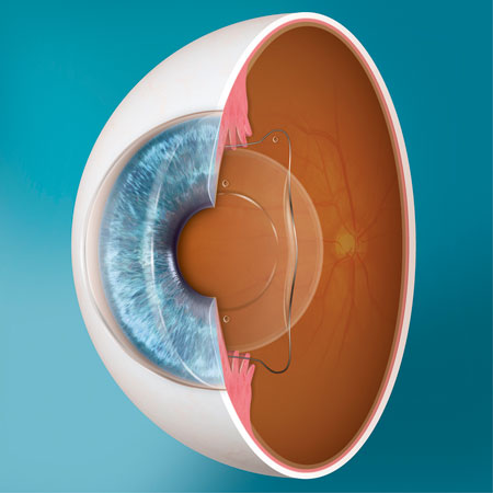
Keratoconus is diagnosed as a common and gradually progressive disease within the eye. The disease is immediately detected when the cornea starts changing its shape irregularly, becoming cone-shaped. This phenomenon is called keratoconus, which leads to various problems.

Causes of Keratoconus: Medical reports suggest that the causes of keratoconus have not been discovered yet. Some opinions indicate that excessive eye rubbing can lead to this disease. Other opinions suggest that it can occur due to prolonged wear of contact lenses. Finally, there is another view confirming that the disease might occur due to genetic factors. There are still different opinions about the causes of keratoconus.

Cost of Keratoconus Surgery: The calculation of the price of keratoconus surgery depends on the needs and requirements of each patient. The price is determined based on the rate and extent of the disease. According to official statements, the cost of keratoconus surgery ranges from 73,878 to 110,818 Egyptian pounds. The more medical attention required, the easier and more successful the surgery will be. The price is calculated based on the presence of the following symptoms:

Symptoms of Keratoconus:

  1. Lack of clear vision.
  2. Experiencing vision problems at night.
  3. Inability to tolerate light, known as light sensitivity or photophobia.
  4. Significant eye fatigue.
  5. Central area damage, making the cornea extremely thin.

Best Doctor for Keratoconus Treatment in Egypt: Dr. Ahmed Al-Motasem for Eyes and Lasik is one of the most skilled specialized doctors in detecting and treating keratoconus. One of the most important factors in the success of this procedure is relying on a highly experienced doctor in this vital area, which is one of the most sensitive parts of a person.

Determining Keratoconus Treatment Options: Dr. Ahmed Al-Motasem first conducts a thorough examination of the cornea to diagnose the condition. The idea of keratoconus surgery can be replaced with the option of wearing contact lenses. However, this option is not highly effective, so opting for surgery might be the optimal solution.

Performing Keratoconus Surgery: When the decision for surgery is made by Dr. Ahmed Al-Motasem, he provides a comprehensive explanation of the procedure. The reason for this is that while the surgery can be highly effective, it may have some complexities, such as the cornea not accepting donor tissues, and a longer recovery period.

Key Alternative Treatments for Keratoconus Surgery: Dr. Ahmed Al-Motasem avoids keratoconus surgery as the last resort, opting for the most recent treatments instead:

  1. Cross-Linking Treatment for the Cornea: Through this treatment, the problem’s progression can be controlled, and it can be used to prevent the rapid spread of the disease.
  2. Implanting Rings for Keratoconus Treatment: These rings can be implanted inside the cornea using laser technology. Dr. Ahmed Al-Motasem implants two half-circle rings of varying thickness. The procedure can be tolerated by the cornea and cannot be rejected. One of the main advantages of this surgery is that it flattens the cornea, providing better vision.
p-STAAR_MICL_Illu_7

Refractive and Cataract Solutions with lences (IOL-ICL)

Refractive Solutions

Myopia, hyperopia and astigmatism are all common vision conditions that affect large numbers of people. Traditionally treated through corrective eyewear or contact lenses, the EVO Visian ICL and Visian ICL family of lenses are designed to provide visual freedom to patients, lessening or eliminating the reliance on glasses or contacts.


EVO Visian ICL™

STAAR® Surgical’s phakic IOL for myopia and astigmatism

EVO is an Evolution in Visual Freedom designed to provide premium refractive outcomes while optimizing patient comfort. Made from Collamer®, EVO can treat a wide range of refractive error. 

EVO+ Visian ICL™

STAAR® Surgical’s phakic IOL for myopia and astigmatism with an expanded optic

EVO+ continues the Evolution in Visual Freedom with an Expanded Optic designed for patients with larger pupils. Also made from Collamer®, EVO+ is based on the proven EVO platform and is the newest generation of EVO lenses.

Visian® ICL™

STAAR® Surgical’s phakic IOL for myopia, hyperopia and astigmatism

The Visian ICL is made from Collamer, a biocompatible material created and used exclusively by STAAR Surgical. This lens is able to treat a large range of refractive error varying by region.

Cataract Solutions

Cataracts are the clouding of the eye’s natural lens, which inhibits light from easily passing through the lens. The clouded lens can be removed and replaced with an intraocular lens (IOL). STAAR® Surgical Company markets various models of Collamer® and Preloaded IOLs. These innovative products are designed to conveniently deliver our high-quality IOLs through a small incision in a predictable, efficient manner.

Collamer IOLs

STAAR® Surgical’s aphakic IOL for cataracts

One of STAAR’s primary Collamer IOLs is the nanoFLEX IOL. nanoFLEX is a single-piece lens designed to treat aphakia. This lens combines STAAR’s proprietary Collamer material with an advanced C2 aspheric optic.

Preloaded IOLs

STAAR® Surgical’s Preloaded IOLs for cataracts STAAR markets preloaded intraocular lenses in certain geographies, with silicone or acrylic optics.  These innovative products are designed to conveniently deliver our high-quality IOLs through a small incision in a predictable, efficient manner.

keratoconus

Keratoconus: how to protect your eyes from a gradual drop of vision

this condition is one of the  most popular chronically or slowly progressive ocular conditions that takes years to affect and impair a person’s vision. In this condition the cornea, the clear window at the front of the eye loses its natural spherical contour, and becomes thinned out ,bulging out at weak points and assuming a conical shape. This abnormality of shape and contour affects the bending of light rays at the corneal interface , causing irregular bending and defocus of these rays when they fall onto the retina, leading to drop of visual clarity and acuity. The condition affects one or both eyes with varying degrees, and the average age of affection is between 15 -25 years . 

What causes keratoconus?

The actual cause is unknown yet, but  research has shed light that a deficiency of certain building enzymes within the corneal stroma.  

The wear and tear that is constantly repaired by these enzymes is slowed down, causing excessive accumulation of diseased collagen fibers and oxygen free radicles, leading to weakening of the corneal stromal bridges, and hence weakness  and forward bulge of the cornea. 

Research has also linked the condition to certain hereditary factors and genetic conditions, which highlights the importance of family history in this condition. 

In addition, excessive unprotected exposure to sunlight and constant eye rubbing have also been implicated. Also, the misuse of poor-quality contact lenses has also been added to the predisposing factors. 

Available treatment options:

At the early stages of the disease , glasses and/or rigid contact lenses (Rose -K, RGP) can be used to treat the condition according to your doctor’s decision. 

As the condition worsens however, your ophthalmologist or eye surgeon may resort to corneal collagen cross linking or intracorneal ring implantation. 

What are the common symptoms for keratoconus?

Patient’s with this condition notice that their vision becomes gradually blurred and unclear with frequent change of their glasses prescription.

 This subtle symptom may progress over years or months and may affect both eyes and one eye more than the other .

 the most common early symptoms include 

  • Blurring of distant and near vision despite wearing the proper optical correction
  • Straight lines appear bent or skewed at different angles
  • Increased sensitivity to ,light and increased sense of haloes around the light 
  • Redness and swelling of the eye

Later symptoms become more ominous and include 

  • Increased blur and visual distortion that does not respond to  any form of optical correction
  • Intolerance to contact lenses
  • Progression of myopia and astigmatism

What are the various surgeries that can be used to treat the condition?

Corneal collagen cross linking:

 this procedure uses ultraviolet light rays and concentrated vit B2 eye drops to create and strengthen the interconnecting bridges between the collagen fibers in the corneal stroma, making it stronger and less liable to bulge. 

The vitamin B2 drops are instilled onto the eye over a period of 30 minutes, this is followed by further 30 minutes of ultraviolet exposure. 

intracorneal ring implantation

 these semicircular devices made of PMMA are inserted into tracks created within the stroma of the diseased cornea and help to restore visual clarity and acuity by flattening the cones and increasing corneal thickness therefore creating a smoother refractive surface and regular light refraction onto the retina.

Corneal implantation

 if the cornea is severely thinned out or damaged you might need to have it partially or fully replaced by  donor tissue. This surgery restores the natural anatomy of the cornea and may provide best visual outcomes for some cases

Not all keratoconus patients have the same condition or follow the same treatment protocol, or  respond to the same treatment modality, each treatment option is specifically tailored according to the presented case 

elmoatassem-lasik-img-1

FemtoLASIK surgery in Egypt

What is the FemtoLASIK operation in Egypt?

The femtoLASIK procedure is different from the normal LASIK procedure in that the treating physician does not use the traditional blades and surgical instruments that are used to create a flap on the outer surface of the cornea, and instead uses two types of lasers, one to create the flap and the other to correct the error.  femtoLASIK procedure in Egypt is also known as the blade-free lasik procedure.

Are there any conditions for femtoLASIK procedure?

Before deciding to perform any operation, your ophthalmologist will first examine your eyes and ask you a series of very important questions  to make sure that you are  ready for the procedure. Similarly, the indications for the procedure are not very different from the conditions for normal LASIK procedure, i.e. every patient that is  eligible for the normal LASIK operation is qualified. Regardless of the patient’s readiness, some medical studies have shown to prefer femtoLASIK over traditional LASIK, to avoid its possible complications as a result of the use of a bladed  microkeratome on the surface of the eye.

What is the mechanism for femtoLASIK?

In the femtoLASIK procedure, the doctor uses a type of ultra rapid laser known as the femtosecond laser to create  a thin outer flap of corneal tissue, then uses another type of laser known as eximer laser  to reshape the corneal tissue under this flap to correct the patient’s eyesight, and then elevated flap is returned to its original state after the corneal tissue is treated beneath it.

Advantages of using femtoLASIK

Recently laser operations in the field of ophthalmology have gained a wide reputation and popularity among doctors and patients alike, and have become one of the most important methods used in the treatment of visual problems and freedom from glasses and contact lenses ,  because  of its multiple features,

The most important of these features are:

  • The high speed and precision of the femtosecond laser, which makes it a safe alternative to microkeratome blades used in normal LASIK operations, and avoids complications of traditional treatments.
  • FemtoLASIK is completely painless, an important feature that is not available in many other therapeutic modalities.
  • Laser technology gives greater control precision, and accuracy than other means.
  • High-speed laser technologies also allow meticulous control of the thickness of the surface corneal flap, providing better chances of correcting vision with never before seen results and accuracy
  • The femtosecond laser allows the doctor to deal with each case with high individuality and custom tailor the treatment according to your needs, giving you the maximal visual results, and does not have to generalize one method of treatment for everyone.
  • Another important feature of femtoLasik is that it absorbs relatively weak corneal fish conditions without fear of bad complications.
  • With the development of laser techniques, the risk of scars on the surface of the cornea or eye exposure to corneal nerve injury and dry eye caused by microkeratomes  has decreased, classifying it as a safer technique.

Tips after the FemtoLASIK procedure

After the procedure, the patient is advised to abide by some recommendations and instructions to complete the success of the operation and achieve the desired results, the most important of which are:

  • Be careful not to expose the eye to dust and wind for at least three days.
  • Be careful not to be exposed to extreme lighting, sunlight and high heat sources.
  • Make sure that water does not enter the eye for at least three days.
  • Be careful not to expose the eye to the radiation of electronic devices and smartphone screens for one to three days after the procedure.
  • Take the recommended treatment from your doctor regularly and seriously as these eyedrops very important for the success of the surgery.

What is the cost of the FemtoLASIK operation in Egypt?

As is known in the medical field in general, the cost of any operation varies greatly according to the hospital fees, the procedure done, materials used, as well as the skill, experience, and knowledge of the treating doctor and his scientific qualifications and the balance of his previous successful operations.
The cost of the Lasik operation

What is the difference between the FemtoLASIK procedure in Egypt and the FemtoSMILE?

FemtoLASIK surgery in Egypt:

As mentioned  earlier, the femtoLASIK procedure is different from the normal LASIK in that the your ophthalmologist does not use the surgical blade in the femtoLASIK procedure to remove the surface flap from the corneal tissue. In this  procedure a type of laser known as the femtosecond laser is used instead. In another type of laser vision correction, the femtoSMILE, the corneal flap is not created or elevated at all, but the femtosecond laser work is done directly without the need to separate corneal layers, and this is the difference between the two procedures femtoSMILE and femtoLASIK.

The LASIK procedure is divided into two main parts:
  • Phase 1: The laser-designed eye phase, by removing a thin flap from the corneal surface of the cornea using a microkeratome blade in normal LASIK operations, and using femtosecond lasers again in femtoLASIK.
  • Phase 2: The next stage of the elevation phase of the surface flap, after which the lasik device is used to adjust the degree of curvature of the cornea and its  thickness, in order to correct the error.

FemtoSMILE surgery

This  modern technique is used in the treatment of myopia in which femtosecond laser  is used to separate a thin part of the corneal stroma called the lenticule and then the doctor removes this part through the pocket incision at the edge of the cornea through a partial thickness incision without the need to raise a flap at all

femtoSMILE is used in the treatment of myopia in the following cases

  • In cases where the thickness of the cornea is lower than necessary to allow lasik or femto lasik operations.
  • femtoSMILE  is also used in cases where myopic errors are too high to be corrected using lasik or femtoLASIK.

When is the femtoSMILE better than the femtoLASIK?

Only the attending physician can determine the optimal or best treatment method for the patient, and in some cases it is better to use femtoSMILE instead of femtoLASIK according to doctor’s experience, knowledge and skill.

The doctor will make the decision to use femtoSMILE instead of femtoLASIK when the following conditions are met:

  • low corneal thickness  .
  • Highly curved cornea.
  • High degree of myopia.
  • Low astigmatism

When the patient’s condition allows the use of femtoLASIK or femtoSMILE evenly, which is better?

In some cases, the possibility of using the two procedures is equivocal: femtolasik and femtosmile, i.e. the patient is ready for any of the two operations according to the results of the examinations and images  performed by the doctor,The cost of the femtoSmile surgery in Egypt

The femtoSMILE procedure 

  • The femtoSMILE is minimally invasive and safe procedure, where the laser machine touches the outer surface of the cornea at a very light degree.
  • The device then separates a very fine slice of the internal corneal tissues called a lenticule , opening a very small incision of about two and a half millimeters at the side of the cornea.
  • The doctor then extracts the thin layer lenticule from the inside of the cornea, corrects the defects of the surface of the cornea and the degree of curvature
  • Immediately after the surgery, the patient’s vision maybe blurred, and the vision gradually returns after the effects of the surgery subside and the patient begins to follow the doctor’s instructions for a safe and successful  recovery plan.

Post-FemtoSMILE

After the femtoSMILE, the doctor gives some recommendations and instructions to the patient, the most important of which are:

  • Rest your eye for up to a week, and do not engage in any stress-prone contact sports  or focused activity, such as reading.
  • Do not expose your eyes to excessively bright lighting, refrain from looking at electronic screens and smartphones, and refrain from watching TV as well, until the doctor allows you to do so after the necessary follow up. 
  • Do not expose the eye to dust, smoke and heat for a week or more as decided by your ophthalmologist.
  • Refrain from using make up powders and cosmetics until the doctor allows the you to do so.
  • Make sure that water does not enter the eye when washing the face or bathing at least during the first week.
  • Do not expose the eye to sunlight, and protect the eye when you leave the house during the day by wearing sunglasses.
The cost of intracorneal ring implants in Egypt

The cost of intracorneal ring implants in Egypt

Treatment of keratoconus with intracorneal rings

The treatment of keratoconus with intracorneal rings is one of the most effective ways to treat the condition, and is done by placing half-circle or full circle segments of rings into tunnels created in the corneal. 

Mechanism of treatment of keratoconus with rings

  • Topical anesthesia is first instilled to numb the eye and prevent any discomfort
  • The rings are implanted at the periphery of the cornea  so that the center of the cornea remains free without being touched. They are implanted in carefully constructed tunnels , which can be either created manually or by femtosecond laser 
  • The rings are inserted into the cornea.
  • They aim at  reducing irregular astigmatism by supporting the base of the cone and flattening the bulge
  • These rings can be removed if necessary, but in such a case, the cornea will return to shape before surgery.

Advantages of the implantation of keratoconus rings

  • The main purpose of treating keratoconus with rings is to restore the shape of the natural eye and allow the patient to tolerate  contact lenses.
  • This type of treatment is fairly good, especially for people who do not want to have a corneal transplant keratoplasty in Egypt.
  • This treatment can improve vision c=and can be accompanied by eyeglasses

What are the steps of the operation?

  • Before starting this procedure, anesthetic eye drops are applied first.
  • After that, the doctor applies the eyelid speculum, which essentially keeps the eye open during the procedure.
  • The doctor creates a tunnel inside the cornea, by laser or manually.
  • The rings are then inserted into the cornea
  • Then, at the end of the procedure, the doctor applies drops and a plastic shield for protection.

Cost of implanting intracorneal rings for  keratoconus in Egypt

In Egypt it is considered a common operation, it varies in cost and depends on several main factors which are the type of tunnel creation procedure used in the operation, the hospital and doctor fees, and the cost of the rings and their type. 

What are the conditions for implanting intracorneal rings?

  1. There should be  no opacity (scar) on the surface of the cornea (cornea must be clear).
  2. The corneal thickness should be within normal limits 

What are the expected results after the procedure?

  • Vision is expected to begin to improve in the week following the procedure by 50%  but may fluctuate for several days until it stabilizes, and most people can return to work one or two days after the treatment.
  • Additional procedures may be used to  complement procedures such as corneal stabilization and other techniques.
  • It may be done more than once with various ring sizes and positions to accommodate the changing corneal parameters .

What are the rings used to treat keratoconus?

implantation of PMMA rings or ring segments, where they act as stents, and  are installed to adjust and stabilize the degree of corneal curvature.

These rings are made up of one or  two semicircular sections, implanted in the cornea, on outside the optical zone, and there are also full segment  rings, the type of ring is chosen according to the condition of each patient. 

These rings are made of solid materials ,PMMA, by installing them in the cornea, they make changes in the shape of the cornea and reduce the bulging cone and provide stability.

What cases require a  corneal transplant?

There are many cases in which corneal transplantation is used in Egypt.

  • Advanced keratoconus. 
  • extreme thinning of the cornea.
  • Scarring or swelling of the cornea, caused by infection or injury.
  • Corneal ulcers and scars , including those caused by infection and not responding to treatment.

It is known that

  • Corneal transplantation contributes to restoring vision, reducing pain, improving the appearance of damaged or sick cornea, or improving its appearance.
  • Most corneal transplants are successful, but corneal transplantation has complications, such as rejection of the donor cornea, so post-operative car and follow up are essential.

The cost of corneal transplantation in Egypt

Results of corneal transplantation

  1. Most people undergoing corneal implants at least partially regain their sight, depending on the cause of the surgery and the patient’s health and eye condition.
  2. Close follow up is needed  because there is a possibility of complications and rejection of the cornea even after years after corneal transplantation, and corneal rejection can often be controlled by medications.
  3. You may need to correct your vision after surgery, and it may take several months for your vision to improve.
  4. When the transplanted  cornea fully heals and recovers – several weeks to several months after surgery, you doctor will make some adjustments that can improve your vision.

Some of these are :

  • Correction astigmatism: meaning that stitches that held the  cornea in place in the patient’s eye can lead to corneal irregularities and astigmatism, causing blurring of vision at some points.

Part of this may be corrected by removing some stitches and tightening others .

  • Correction of other vision problems:  Refractive errors, such as myopia hyperopia can be corrected by wearing glasses, contact lenses or laser eye surgery in some cases.

What is the cost of intracorneal ring implants in Egypt?

The cost of intracorneal ring implant in Egypt using the femtosecond laser device also varies according to the doctor’s skill, experience , knowledge. In addition to the  specifications of rings used in the surgery and whether they will be inserted manually or by Femtolaser which is currently used in Egypt and  is considered to have guaranteed and safe results.

Best Eye Surgeon in Egypt

A lot of skill knowledge and experience is needed and is imperative  for the diagnosis of keratoconus in its early stages  and its modalities of treatment, including the type, placement and thickness of the rings, and whether additional surgeries will be done. The best option for you is to look for the best eye surgeon ophthalmologist  specialized in keratoconus surgeries known for their efficiency and guaranteed results.

 Hence comes Dr. Ashraf Hassan Soliman consultant of keratoconus and corneal surgery, cataract and glaucoma  surgery, refractive surgery and laser vision correction on the top of the best ophthalmologists and  as the best doctor for the treatment of keratoconus in Egypt.

1018_Uveitis

What is uveitis?

What is uveitis?

Inflammation of the line lining of the eye


What are its symptoms?

  • Pain and redness in the eye
  • Redness of the eyes may not appear in children.
  • Blurry vision.
  • Sensitivity to light.
  • Shadows, interlacing lines, or points that float in the field of view

How dangerous is it?

There are types:

  • An acute anterior is one of the simplest types and one of the lowest in terms of complications.
  • Chronic posterior may cause serious complications if not treated.
  • Medium and medium severity.

What are the causes?

Most of the time it is due to an imbalance in the immune system, but sometimes the cause is a microbe.

Strabismus-678x446

The evolution of strabismus surgery

An exciting development has occurred in strabismus surgeries in recent years, including, for example, strabismus surgeries without the use of any surgical sutures, which prevents any problems during surgery and the surgery is completely safe, as well as the patient does not feel any inconveniences after surgery and returns to his activities immediately.

It is also possible to perform strabismus surgeries using surface anesthesia drops and not to resort to general or local anesthesia, which increases the accuracy of controlling the angles of the squints using modern surgical methods with which the patient does not feel any pain, which increases the accuracy and success of the surgical procedure.

Note that strabismus surgeries are considered one-day surgeries, and the eye is not covered after the operation, and the patient returns to his activities immediately after surgery.

Multifocal lenses2

Multifocal lenses treat cataracts on eyes

With the proliferation of electronic devices and cell phones, the eye problems associated with them have increased. With age, the natural lens of the eye loses its ability to clarify the image, which is known as white water on the eyes, but it has become accompanied by other visual defects such as myopia, farsightedness and medial vision.

In the past, the only options available to patients were either monofocal or bifocal lenses, but eyeglasses are still often required for certain activities. With the development of modern technologies, one of the most recent methods of treating cataract on the eyes is Trifocal IOL.

What are multifocal lenses for treating white water on eyes?

Trifocal lenses are the most advanced lenses for treating cataracts, as they are implanted in the eye during a cataract or lens replacement procedure.

It is a bifocal lens (Bifocal), but it was designed with the addition of a different feature in it that makes the main goal of it to provide a clear vision of the near, medium and far distances for the patient with cataract on the eyes.

What is the difference between multifocal lenses and other lenses for treating cataracts?

1- Monofocal lenses.

The most common type of lens used to treat cataracts. Unlike your natural lens, this lens features a single fixed distance focus. It means treating patients with farsightedness only, but after the operation you will need glasses for reading or watching closely.

2- Bifocal lenses.

Like bifocal eyeglasses, this one has areas that help you see things at different distances (far and near only). It may take several months for your mind to adjust for your vision to appear normal.

3- Multifocal Lenses.

Trifocal lenses work by gathering light entering the eye from near, medium, and far distances, focusing it on a single focal point on the retina. As a result, it can see objects at various distances, providing close, middle and far vision through a single lens.

This type of lens is the latest in the world of intraocular lens implantation, and it substitutes for the use of eyeglasses.

Toric IOLs are the latest in multifocal lenses

Toric lenses are eye lenses that correct astigmatism as well as nearsightedness or farsightedness.

Immediately before cataract surgery, the surgeon places temporary markers on the patient’s cornea that determine the location of the most curved meridian at the front of the eye, and then implantation of the (Toric IOL) lens during cataract surgery is performed on the eye, the surgeon rotates the lens so that the existing marks are aligned. On the lens with marks on the cornea to ensure proper astigmatism correction.

What are the advantages of multifocal lenses for treating white water on eyes?

Unlike monofocal and bifocal lenses, the design of multifocal lenses includes a third focal point (middle distance) as well as near and far distances; This is what a person requires to do daily activities such as:

  1. Reading.
  2. Cooking, cleaning and ironing.
  3. Work on the computer.
  4. Shopping.
  5. Watching TV.
  6. Leadership.
  7. Work in the garden.
  8. Doing sports.

What are the disadvantages of multifocal lens implantation for cataract treatment?

Multifocal implantation, like many modern technologies, suffers from some disadvantages, which is that patients suffer from the appearance of some bright rings around lights at night. While the patient does not see it during the day. Therefore, if you are a practitioner of night professions, Prof. Ahmed Al-Mu’tasim advises that you consult your doctor about the compatibility of the process with your profession and your daily routine.

What to expect after implantation of multifocal lenses?

After lens implant surgery, patients stay in the hospital under observation for one or a few nights. And with the use of modern surgical methods; The patient can leave the hospital on the same day. The patient is instructed on how to use eye drops that contain antibiotics to prevent infection, which he should use for 3 weeks after the operation. The patient is provided with eye drops to prevent eye infection.

Who are the candidates for multifocal lenses?

  • People over the age of 55 who rely heavily on glasses.
  • Astigmatism patients.
  • Patients with vision disorders such as myopia or hyperopia.
  • Those with age-related eye diseases such as cataracts.

However, not all candidates are suitable for all types of lenses, for example; Sometimes, the best visible results are achieved after a cataract operation with a different type of lens.

For example, you may have astigmatism in one eye than in the other. In this case, your eye surgeon recommends using an IOL lens in that eye only.

An eye surgeon can fully evaluate your specific needs during a pre-operative exam, and help you choose the best type of lens for a successful result and better vision.